EKG Translated — Preview Edition — Daisy Rice
⚠️ AUTHORIZED USE ONLY — NOT FOR DISTRIBUTION ⚠️
Licensed copy. All rights reserved.
Preview Edition • A Guide for Clinicians & First Responders
EKG
Translated
A Preview of Understanding STEMI, Occlusion MI,
and the ECG Patterns That Save Heart Muscle
Author
Daisy Rice
2025 • Preview Edition
⚠️ AUTHORIZED USE ONLY — NOT FOR DISTRIBUTION ⚠️
Licensed copy. All rights reserved.
Preview Edition • A Guide for Clinicians & First Responders

EKG
Translated

A Preview of Understanding STEMI, Occlusion MI, and the ECG Patterns That Save Heart Muscle

⚠️ AUTHORIZED USE ONLY — NOT FOR DISTRIBUTION ⚠️
Licensed copy. All rights reserved.
ii

“Time is muscle.”

— Ancient Cardiology Proverb

⚠️ AUTHORIZED USE ONLY — NOT FOR DISTRIBUTION ⚠️
Licensed copy. All rights reserved.
EKG Translated Overview
iii

Preview Overview

This preview of EKG Translated introduces two critical concepts in emergency cardiology: the classic ST-elevation myocardial infarction (STEMI) and the emerging paradigm of Occlusion MI (OMI). Its sample chapters cover characteristic ECG patterns, STEMI equivalents such as posterior MI, de Winter pattern, Wellens syndrome, and Sgarbossa criteria for left bundle branch block. It also discusses the role of AI in detecting subtle ischemic changes and provides a curated list of resources for further learning.

The full book will include hundreds of annotated ECGs, video tutorials, detailed treatment protocols, and interactive quizzes to build rapid interpretation skills. This preview offers a glimpse of the core framework that the complete edition develops in depth.

Keywords: STEMI · Occlusion MI · de Winter · Wellens · Sgarbossa · ECG · Posterior MI · AI · Emergency Cardiology · Preview

⚠️ AUTHORIZED USE ONLY — NOT FOR DISTRIBUTION ⚠️
Licensed copy. All rights reserved.
EKG Translated Contents
iv

Preview Table of Contents

Chapter One
Chapter Three
Chapter Four
References

The complete book contains 12 additional chapters,
hundreds of ECG tracings, and interactive case studies.

⚠️ AUTHORIZED USE ONLY — NOT FOR DISTRIBUTION ⚠️
Licensed copy. All rights reserved.
I

Chapter One

Understanding STEMI

The Most Dangerous Type of Heart Attack and the ECG Patterns That Identify It

⚠️ AUTHORIZED USE ONLY — NOT FOR DISTRIBUTION ⚠️
Licensed copy. All rights reserved.
EKG Translated Understanding STEMI
1

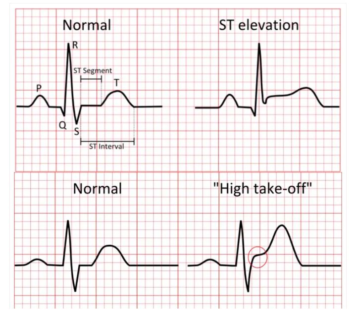
A STEMI, or ST-elevation myocardial infarction, is the most dangerous type of heart attack. It occurs when a coronary artery becomes completely blocked—usually after a cholesterol plaque ruptures and a clot rapidly forms—cutting off oxygen to a section of the heart muscle. On a 12-lead ECG, this appears as an abnormal upward lifting of the normally flat ST segment above the baseline, creating a shape some clinicians compare to a tombstone. This elevation is visible in contiguous leads that look at the same part of the heart (such as the inferior, anterior, or lateral walls), signaling full-thickness injury. The BMJ Best Practice guideline defines STEMI as new or increased, persistent ST-segment elevation in at least two contiguous leads of ≥1 mm in all leads other than V2–V3, where more specific age- and sex-based cutoffs apply: ≥2.5 mm in men under 40, ≥2 mm in men over 40, and ≥1.5 mm in women regardless of age.

Typical symptoms include crushing chest pain that may radiate to the jaw, arm, or back, along with shortness of breath, sweating, nausea, or lightheadedness. However, women, older adults, and people with diabetes often experience more subtle signs—what clinicians call anginal equivalents—such as dyspnea, fatigue, syncope, or persistent vomiting. Because "time is muscle," meaning every minute of delay causes more permanent heart damage, emergency treatment focuses on reopening the artery as fast as possible.

⚠️ AUTHORIZED USE ONLY — NOT FOR DISTRIBUTION ⚠️
Licensed copy. All rights reserved.
EKG Translated Understanding STEMI
2

The preferred method of reperfusion is percutaneous coronary intervention (PCI), which uses a balloon and stent to restore blood flow. If a catheterization lab is not immediately available, fibrinolytic drugs may be given as a bridge. Additional standard medications include aspirin, anticoagulants, statins, and beta-blockers. A rise in cardiac-specific troponins confirms the diagnosis, but treatment should never be delayed while waiting for laboratory results.

If you or someone nearby develops symptoms that could be a STEMI, call emergency services immediately—do not drive yourself to the hospital—because rapid intervention is the single most important factor in saving heart muscle and improving survival. Emergency medical services can acquire and transmit a pre-hospital ECG, activate the cath lab from the field, and begin treatment en route. As the SAEM M3 Curriculum emphasizes, patients who present to the emergency department with chest pain should have an ECG obtained and interpreted within 10 minutes of arrival.

· · ·
⚠️ AUTHORIZED USE ONLY — NOT FOR DISTRIBUTION ⚠️
Licensed copy. All rights reserved.
EKG Translated ECG Reference Image
3

Figure 1 — STEMI ECG Pattern

STEMI ECG

[Image could not be loaded. Please verify the image URL and reopen with an internet connection.]

ST-segment elevation in contiguous leads. Note the characteristic "tombstone" morphology.

⚠️ AUTHORIZED USE ONLY — NOT FOR DISTRIBUTION ⚠️
Licensed copy. All rights reserved.
EKG Translated Understanding STEMI
4

The classic STEMI presentation is now understood to be just one manifestation of a broader spectrum of acute coronary occlusion. While the "tombstone" ST elevation is the most widely recognized ECG finding, it is far from the only one. The 2025 Australian Clinical Guideline for Diagnosing and Managing Acute Coronary Syndromes has formally introduced the terminology of Acute Coronary Occlusion Myocardial Infarction (ACOMI), recommending broader recognition of ECG patterns beyond ST-segment elevation alone.

Understanding which leads show ST elevation also tells you which coronary artery is likely blocked. Inferior STEMI (leads II, III, aVF) typically involves the right coronary artery or left circumflex. Anterior STEMI (leads V1–V4) usually indicates left anterior descending artery occlusion—the so-called "widowmaker." Lateral STEMI (leads I, aVL, V5, V6) often involves the left circumflex artery. Recognizing these patterns helps anticipate complications and guides emergent management decisions. But as the next chapter explains, not every dangerous occlusion announces itself with ST elevation.

⚠️ AUTHORIZED USE ONLY — NOT FOR DISTRIBUTION ⚠️
Licensed copy. All rights reserved.
II

Chapter Two

Beyond STEMI: The Occlusion MI Paradigm

Why a Normal-Looking ST Segment Does Not Rule Out a Major Heart Attack

⚠️ AUTHORIZED USE ONLY — NOT FOR DISTRIBUTION ⚠️
Licensed copy. All rights reserved.
EKG Translated The Occlusion MI Paradigm
5

The traditional approach to heart attacks has long been to classify them as either STEMI (with clear ST elevation on an ECG, presumed to be a complete blockage) or NSTEMI (without such elevation, often considered a partial blockage). However, cardiology is now undergoing a major shift because many patients with a completely occluded coronary artery—and ongoing, extensive heart muscle death—never show classic ST elevation on their ECG. Research has shown that more than half of patients with an occluded artery will not show ST-segment elevation, and this can lead to delays in care and worse outcomes.

This realization has given rise to the concept of Occlusion MI (OMI), which asks not "Does the ECG have ST elevation?" but rather "Is there an acutely occluded artery?" The OMI paradigm shifts the focus from rigid ECG thresholds to a pathophysiology-based strategy that integrates clinical assessment, ECG interpretation, and adjunct diagnostic modalities to actively seek evidence of acute coronary occlusion. As Stephen Smith, MD, a leading proponent of this paradigm, has stated: "We think we should get rid of the term ST-elevation MI, because it focuses on ST-elevation and makes you think that ST-elevation is all that matters—when what matters is acute coronary occlusion without collateral circulation leading to imminent myocardial necrosis or death."

⚠️ AUTHORIZED USE ONLY — NOT FOR DISTRIBUTION ⚠️
Licensed copy. All rights reserved.
EKG Translated The Occlusion MI Paradigm
6

Several so-called STEMI equivalents can indicate a dangerous occlusion even when the ST segment is not elevated. These patterns require the same urgency as classic STEMI because they signify the same underlying pathology: an acutely occluded coronary artery causing ongoing myocardial necrosis. Missing these patterns leads to delays in reopening the artery, which directly costs heart muscle. Studies confirm that STEMI criteria are a poor surrogate marker for acute coronary occlusion, and that OMI signs have approximately double the sensitivity with preserved high specificity. The key STEMI equivalents include:

Posterior MI. When the back wall of the heart is infarcted, the standard 12-lead ECG may show only ST depression in leads V1–V3 rather than elevation anywhere. Deep, horizontal ST depression in V1–V3, often accompanied by tall R waves and upright T waves, should raise immediate suspicion. Posterior leads (V7–V9) can confirm the diagnosis by revealing ST elevation ≥0.5 mm, but treatment should not be delayed.

De Winter Pattern. First described in 2008 in the New England Journal of Medicine, this pattern is associated with proximal LAD occlusion. It is characterized by 1–3 mm of upsloping ST depression at the J-point in precordial leads (typically V1–V4), which continues into tall, symmetric T waves. There is often 1–2 mm of ST elevation in lead aVR. This is considered a STEMI equivalent requiring emergent catheterization.

⚠️ AUTHORIZED USE ONLY — NOT FOR DISTRIBUTION ⚠️
Licensed copy. All rights reserved.
EKG Translated The Occlusion MI Paradigm
7

Wellens Syndrome. This pattern is associated with critical stenosis of the proximal LAD, characterized by deeply inverted or biphasic T waves in V2–V3, which may extend to V1–V6. Critically, these changes typically appear when the patient is pain-free. Wellens syndrome carries up to 99% specificity for significant LAD stenosis.

Sgarbossa Criteria for Left Bundle Branch Block. Diagnosing acute MI in the presence of LBBB is notoriously difficult. The Sgarbossa criteria and the Smith-modified Sgarbossa criteria (using the ST/S ratio) provide validated methods for detecting acute occlusion in patients with LBBB or ventricular paced rhythms.

⚠️ AUTHORIZED USE ONLY — NOT FOR DISTRIBUTION ⚠️
Licensed copy. All rights reserved.
EKG Translated The Occlusion MI Paradigm
8

To help address the challenge of detecting these subtle patterns, artificial intelligence tools such as PMcardio are being developed. The PMcardio AI-ECG model (also known as the "Queen of Hearts") has been trained on millions of ECGs to detect occlusive myocardial infarction patterns that may be missed by conventional STEMI criteria. In one study, the AI model outperformed STEMI criteria in terms of accuracy (90.9% vs 83.6%) and sensitivity (80.6% vs 32.5%), with similar specificity (93.7% vs 97.7%).

Some experts express concern about false positives and unnecessary cath lab activations, but the goal is to augment clinical judgment, not replace it. As Smith notes, "It can see these waveform differences, just like it can recognize a face. It can recognize OMI on an EKG."

Practical Takeaway: A normal-looking ST segment does not rule out a major heart attack. If a patient has concerning symptoms and the ECG shows any of these higher-risk patterns, do not wait for textbook ST elevation. Emergently consult cardiology, consider activating the cath lab, and treat the patient as having a possible occlusion MI.

⚠️ AUTHORIZED USE ONLY — NOT FOR DISTRIBUTION ⚠️
Licensed copy. All rights reserved.
III

Chapter Three

ECG Image Reference

Visual Reference for ST-Segment Elevation and Reciprocal Changes

⚠️ AUTHORIZED USE ONLY — NOT FOR DISTRIBUTION ⚠️
Licensed copy. All rights reserved.
EKG Translated ECG Reference
9

Figure 1 — Diagnostic ECG Pattern

ECG showing STEMI pattern

[Image could not be loaded.]

Review this tracing for ST-segment morphology, reciprocal depression, and the distribution of changes across contiguous leads.

⚠️ AUTHORIZED USE ONLY — NOT FOR DISTRIBUTION ⚠️
Licensed copy. All rights reserved.
IV

Chapter Four

For Further Learning

Curated Resources for Deeper Training in ST Elevation and ECG Interpretation

⚠️ AUTHORIZED USE ONLY — NOT FOR DISTRIBUTION ⚠️
Licensed copy. All rights reserved.
EKG Translated Further Learning
10

The ECG patterns covered in this preview represent a foundation. Continued education and exposure to tracings are essential for developing the pattern-recognition skills that distinguish expert interpreters. The following resources offer case-based learning, detailed explanations, and additional practice with both classic STEMI presentations and the more subtle STEMI equivalents.

Recommended External Resources

The complete book provides dozens more resources, video tutorials, and interactive ECG quizzes.

⚠️ AUTHORIZED USE ONLY — NOT FOR DISTRIBUTION ⚠️
Licensed copy. All rights reserved.

References

Literature & Sources Cited

⚠️ AUTHORIZED USE ONLY — NOT FOR DISTRIBUTION ⚠️
Licensed copy. All rights reserved.
EKG Translated References
11

1. BMJ Best Practice. ST-elevation myocardial infarction. Updated March 2025. https://bestpractice.bmj.com/topics/en-gb/150

2. Brieger DB, Cullen LA, Briffa TG, et al. National Heart Foundation of Australia and Cardiac Society of Australia and New Zealand: Australian Clinical Guideline for Diagnosing and Managing Acute Coronary Syndromes 2025. Med J Aust 2026;224(2). doi:10.5694/mja2.70127

3. Baranchuk A, Bayés-Genis A. Naming and classifying old and new ECG phenomena. CMAJ 2016;188(7):485–486. doi:10.1503/cmaj.151209

4. Webb SR. Wellens Syndrome Important to Consider in Chest Pain: Key Points. American College of Cardiology, January 2023. https://www.acc.org/Latest-in-Cardiology/ten-points-to-remember/2023/01/20/17/59/wellens-syndrome

5. Schaafsma AE, Coolsma C, Lameijer H. Left bundle, right diagnosis. Neth Heart J 2019;27(4):218–221. doi:10.1007/s12471-019-1256-z

6. Sgarbossa EB, Pinski SL, Barbagelata A, et al. Electrocardiographic diagnosis of evolving acute myocardial infarction in the presence of left bundle-branch block. N Engl J Med 1996;334(8):481–487.

7. Smith SW, Dodd KW, Henry TD, et al. Validation of the Modified Sgarbossa Criteria for Acute Coronary Occlusion in the Setting of Left Bundle Branch Block. Am Heart J 2015. Reviewed at: https://coreem.net/journal-reviews/modified-sgarbossa-criteria/

8. O'Riordan M. Should We Be Looking for 'Occlusion MI' Rather Than STEMI? TCTMD, May 2025. https://www.tctmd.com/news/should-we-be-looking-occlusion-mi-rather-stemi

⚠️ AUTHORIZED USE ONLY — NOT FOR DISTRIBUTION ⚠️
Licensed copy. All rights reserved.
EKG Translated References
12

9. Kobo O, et al. From ST-Segment Elevation MI to Occlusion MI: The New Paradigm Shift in Acute Myocardial Infarction. JACC Adv 2024. doi:10.1016/j.jacadv.2024.101314

10. External validation of the PMcardio AI-ECG model for detecting occlusion myocardial infarction in a Portuguese cohort. Eur Heart J - Digital Health , January 2026. Presented at ESC 365.

11. de Winter RJ, Verouden NJW, Wellens HJJ, Wilde AAM. A new ECG sign of proximal LAD occlusion. N Engl J Med 2008;359(19):2071–2073.

12. Maimonides Emergency Medicine Residency. STEMI Equivalents. https://www.maimonidesem.org

13. Sekhon N, Gallegos M. STEMI — Electrocardiogram — M3 Curriculum. Society for Academic Emergency Medicine, updated 2023. https://www.saem.org

14. Potter T, Spencer K, White MD, et al. A 56-Year-Old Female With Acute ST-Segment Elevation Myocardial Infarction, Complete Heart Block, and Hemodynamic Instability. Cureus 2021;13(1):e12857. doi:10.7759/cureus.12857

15. JACC. A New Time Clock for ST-Segment Elevation Myocardial Infarction. J Am Coll Cardiol 2015.

16. Acute ST-Elevation Myocardial Infarction Presenting With Persistent Vomiting. Cureus 2022. PMC.

17. PMcardio for Individuals. Powerful Medical. https://www.powerfulmedical.com